The following is a review of the first 32 cases of partial knee replacement at Peace Arch Hospital (December 2000 to February 2002). It describes the process of implementing a new procedure into an existing surgical practice and into an existing surgical facility.
Partial knee replacement (also called unicompartmental knee replacement, uni-knee or sometimes the 'Oxford Knee') is not a new concept. This method of replacing a small part of the knee has been studied and performed since the 1970's. The early results in North America were not encouraging. Failure rates five or six years after surgery were unacceptably high. Furthermore, this procedure initially required as large an incision and was as traumatic as for a total knee replacement. However, in 1999, I reviewed the work of Murray, Goodfellow and O'Connor -- they showed 98 percent survival of the Oxford partial knee replacement at 10 years. Obviously, significant steps had been made in combating the early difficulties.
By giving particular care to patient selection, the type of artificial component used, and surgical technique, partial knee replacement was successfully introduced into orthopaedic practice at the Peace Arch Hospital.
When I started general orthopedic practice in September 1999, I quickly realized that the main focus of my workload would be treatment of osteoarthritis (wear and tear on the joints). I encountered a significant number of people with severe functional restrictions due to wear and tear on one side of the knee (unicompartmental osteoarthrosis). The standard surgical treatment for these people was total knee replacement or, much more controversial, high tibial osteotomy. In the spring of 2000 I started evaluating younger, potentially more active, patients (usually age 50 or less) for suitability for high tibial osteotomy and by the summer I had several patients on my waiting list.
Around this time, I became aware of the availability of the Oxford Knee in Canada, as well as more recently developed less invasive instrumentation and surgical techniques. After reviewing the pertinent literature, I asked my patients waiting for a high tibial osteotomy, to hold their final decision until I had attended a course in Oxford, England, November 2000.
As it turned out, all of these people changed their mind and wished to be treated with a partial knee replacement because of the advantages it has over high tibial osteotomy:
- the quicker initial recovery, particularly the ability to start weight-bearing as tolerated immediately, rather than staying off their feet for 6-12 weeks until the osteotomy would have healed.
- the possibility of this being a definitive procedure, rather than a procedure that would be expected to lead to total knee replacement in 7-12 years.
- if a later conversion to a total knee replacement would be necessary, the procedure would be expected to be more similar to a first-time total knee replacement than after a high tibial osteotomy.
- the magnitude of surgery is similar for both a unicompartmental knee replacement and a high tibial osteotomy, the risks would be similar in magnitude and frequency as well.
It also has advantages over total knee replacement:
- shorter operating time.
- shorter length of stay.
- shorter initial recovery time.
- less blood loss (meaning a very low likelihood of requiring a blood transfusion).
- the eventual level of function achieved is higher.
|
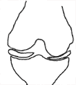 |
| Partially obliterated joint space in one compartment |
Patients considered for this procedure were affected by unicompartmental knee osteoarthrosis, almost always of the medial compartment. X-rays of the knee while bearing weight were obtained for all patients, to assess the narrowing of the affected compartment. If it had not narrowed to be effectively bone on bone (obliteration of joint space), and only partial narrowing could be demonstrated, the patient would be counseled to consider physical activity and conditioning, bracing or high tibial osteotomy.
|
| |
|
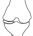 |
| Joint space obliterated in one compartment |
If the joint space was completely obliterated, with preservation of the compartment on the opposite side, the options presented to the patient were bracing, high tibial osteotomy, total knee replacement or unicompartmental knee replacement. The partial knee replacement was presented as an option but was described as a new procedure to North America, with only limited local experience. It was emphasized that the data available to assess this procedure originated from the designer of the procedure (Oxford Group), but also that a more independent evaluation was available from the Swedish joint registry, which confirmed the findings of the Oxford Group. Longevity of the prosthesis was quoted at approximately 95 percent survival of the prosthesis at 10 years.
|
We went through careful discussions to ensure a thorough understanding of the options and if the patient was interested in the partial knee replacement, arrangements would be made to assess the integrity of the ACL. It has been well established by the Oxford Group that ACL integrity is crucial for long-term success. Clinical assessment of the ACL in the osteoarthritic knee is unreliable. More than a simple exam is required. The following strategies can be employed:
- assessment at the time of surgery: lets you proceed with partial knee if the ACL is good or do a total knee replacement if a deficient ACL is encountered. Advantages of this approach include avoiding further expenditure of resources for diagnosis. Disadvantages include the need for dual set up, and the inability to correct the ACL problems and later continue with unicompartmental knee replacement, as may be appropriate for the younger patient. I find this strategy unacceptable, and I have not employed this.
- preoperative MRI assessment. This will visualize the ACL, but will not allow assessment of its functional status.
- scrutinizing the lateral x-ray view can give an indication of the status of the ACL. If clear erosion is seen, which does not completely extend to the posterior aspect of the joint, it is most likely that the ACL is still structurally intact.
- preoperative arthroscopy. This strategy allows inspection of the ACL by seeing its integrity and testing to see how it behaves if the joint is repositioned (hanging loose or still contracting well like an elastic). During this procedure the lateral compartment would be scrutinized for minor problems, which could then be dealt with arthroscopically in preparation for increased load bearing after surgery. It must be emphasized that some wearing of the lateral compartment is not a contraindication to this procedure, as has been well established by the Oxford Group.
Of note, on occasion, no further ACL evaluation was undertaken. Especially with older patients who had other significant health problems. In their case, obtaining further information regarding the ACL would not change the choice of procedure. Even with a deficient ACL, the Oxford knee would be expected to give good benefit for 5 to 6 years, possibly longer for the less active patient.
After suitability for unicompartmental knee replacement was established through arthroscopic evaluation of the knee or MRI, I felt compelled to try and perform this operation within three to four months. It must be emphasized that no clear guidelines are available for this. It is my feeling that if significant more time is allowed to go by, a new evaluation should be undertaken.
Introduction of unicompartmental knee replacement into surgical practice.
This procedure was introduced into Peace Arch hospital in December 2000.
I decided to slot this procedure into the care path that had been developed for total knee replacements. At first we made no changes to the pain management protocol in place for total knee replacements, preferentially using spinal anesthesia with morphine added ('spinal-epimorph') and then allow patients to recover with the use of patient controlled analgesia (pain-killers). This protocol precluded same-day discharge, as overnight monitoring is required after this type of spinal anesthesia. After success with this established method, we are moving to use the rapid recovery protocol that has been developed by the Australian group, Kohan & Kerr. This will allow patients to return home the same day that they have surgery.
Criteria for returning home:
- adequate pain control with oral pain killers
- adequate mobilization to perform activities of daily living safely and with confidence.
- adequate social support.
- satisfactory wound status
These criteria were no different than discharge criteria after total knee or total hip replacement. The patients were instructed to contact my office or Peace Arch Hospital emergency room (evenings and weekends) if any problems would occur. The staples were removed by me at the two week mark. No physiotherapy was initiated in the first two weeks. An individualized decision regarding the use of physiotherapy was made at the two week mark.
Further follow-up was scheduled at two months, six months, and one year.
In analyzing these first 32 cases, data came from the chart, and include age, gender, date of surgery, length of operating room set up and length of surgery, preoperative and postoperative hemoglobin (next day), length of stay, and range of motion at two months and at six months. The patients provided a subjective assessment based on the patient's ability to deal with this process and procedure, both emotionally and physically, looking at postoperative function and postoperative discomfort.
Age:
The average age was 70.19 yr., the youngest patient was 49, the oldest was 84. Eight patients were younger than 65.
Length of Stay:
the length of stay greatly decreased as familiarity with this procedure increased. I speculate that part of this decrease in length of stay is due to my increasing confidence that patients are ready to go home within 24 hours after this procedure, and that this is transferred to the patient during the pre-operative discussions. Once this expectation is set in a patient's mind, the likelihood of the patient feeling ready to recover at home is markedly increased. Furthermore, the length of surgery decreased as well as experience grew, perhaps decreasing the surgical insult, and the consequent need for pain control measures.
Change in Hemoglobin:
No patient required a blood transfusion. From a practical point of view, if a reasonable starting hemoglobin is present, the risk of transfusion is very low.
Length of surgery:
both the length of surgery and its variability decreased as experience grew. Currently, surgical time is usually between 75 and 90 minutes. The decrease in surgical time is mainly due to enhanced experience with the approach and the technical aspects of the operation. A routine has been established and familiarity with the equipment and the procedure has been widely distributed among the operating room nursing staff.
Results for the knee:
Range of motion at two months: 116.00 degrees
Range of motion at six months: 127.14 degrees
| Subjective grading (31 knees): |
| Excellent: |
20 |
| Good: |
6 |
| Fair: |
5 |
| Follow-up over 6 months (14 knees): |
| Excellent: |
9 |
| Good: |
4 |
| Fair: |
1 |
Seeing patients able to move about and flex the joint is very encouraging, especially since this allows them to perform so many daily tasks like climbing stairs or getting in and out of cars. Some discomfort, which is described as different from the original arthritic pain, is common over the medial side of the knee and over the edge of the tibial component. The experience in Oxford indicates that the majority of patients will experience resolution of this discomfort by six months and so far this is the same as my own findings. It is also clear that some patients resume a much higher level of physical activity than typically seen early after total knee replacement. This accounts for some discomfort and swelling, particularly early on. I have now taken the approach to slow people down somewhat, as opposed to encourage them to explore the limits of their ability, as is my practice after total knee replacement.
Conclusion:
- the unicompartmental Oxford knee replacement, utilizing a mobile bearing, has been successfully introduced into my surgical practice and into Peace Arch hospital's orthopedic program
- the condition for which it is designed, unicompartmental osteoarthrosis of the knee, has a high prevalence in our population.
- it has advantages over total knee replacement: shorter operating time, shorter length of stay, less blood loss with a very low likelihood of requiring a blood transfusion. The initial recovery is much quicker, and the eventual level of function achieved is higher. This had already been shown by the Oxford Group, my impression is similar.
- in order to maximize utilization of this technology, it must be understood that partial knee replacement represents a step in a progressive process. If left alone long enough, the wear on the knee will progress and the window of opportunity for a limited procedure, such as unicompartmental knee replacement, will close.
- With significant challenges to management of the resources available in medical practice today, partial knee replacement has potential. It is meant to be a definitive procedure, it increases function rapidly, it allows ready revision if needed, and has limited impact on surgical facilities. However, it is difficult in the current administrative model to transfer savings from one aspect of the medical system to another. This relates mainly to the implant cost, which is approximately 50 percent higher than the currently used total knee replacement system.
- this procedure lends itself for application in stand-alone surgical facilities, with or without overnight stay capability, depending on the choices of anesthetic.